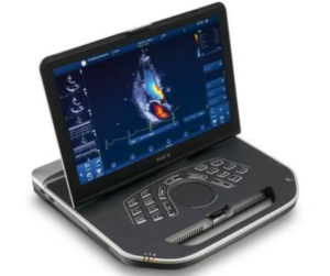
The GE Voluson i has great imaging while covering a wide range of clinical applications. This machine provides you with you with your most demanding needs in a convenient lightweight (12.3 lbs/5.7 kg) laptop design. The GE Voluson i ultrasound machine can be easily used in fixed exam rooms and also emergency and point-of-care applications.
The Voluson i comes with many advanced features for a portable ultrasound.
Imaging Features Include:
Speckle Reduction Imaging (SRI) and CrossXBeam
HD-Flow
Volume Contrast Imaging (VCI)
3D/4D Live 4D Imaging
Tomographic Ultrasound Imaging (TUI)
Virtual Organ Computer-aided Analysis (VOCAL)
Spatio-Temporal Image Correlation (STIC)
SonoVCADheart
SonoNT
SonoVCADlabor
SonoAVCfollicle
SonoRender Start
15″ LCD Screen
Automatic Tissue Optimization (ATO)
Optimized Tissue Imaging (OTI)
Coded Harmonic Imaging
VCI: Volume Contrast Imaging on the Voluson-I utilizes 4D transducers to automatically scan multiple adjacent slices and delivers a real-time display of the ROI. This image results from a special rendering mode consisting of texture and transparency information. VCI improves the contrast resolution and therefore facilitates finding diffuse lesions.
TUI: Tomographic Ultrasound Imaging is a new visualization mode for the Voluson-i in 3D and 4D data sets on the Voluson-i. The data is presented as slices through the data set which are parallel to each other. An overview image, which is orthogonal to the parallel slices, shows which parts of the volume are displayed in the parallel planes. This method of visualization is consistent with the way other medical systems such as CT or MRI, present the data to the user. The distance between the different planes can be adjusted to the requirements of the given data set. In addition it is possible to set the number of planes. The planes and the overview image can also be printed to a DICOM printer, for easier comparison of the ultrasound data with CT and/or MRI data.
SonoVCAD Heart: A technology that automatically generates a number of views of the fetal heart to make diagnosis easier.
SonoAVC follicle: A Voluson-i technology that automatically detects follicles in a volume of an organ (e.g., ovary) and analyzes their shape and volume. From the calculated volume an average diameter can be calculated. It also lists objects according to their size.
SonoVCAD labor: Allows the Voluson-i user to measure fetal progression during the second stage of labor such as fetal head progression, rotation and direction. Visual evidence and objective data of the labor process are provided. All SonoVCAD labor measurements are automatically added to the worksheet
STIC: Spatiotemporal Imaging Correlation is a fetal echo provided by the Voluson-i that visualizes the fetal heart or an artery in static 3D.
VOCAL II: Virtual Organ Computer-aided Analysis is an imaging program on the Voluson-i for cancer diagnosis, therapy planning and follow-up therapy control. It offers contour detection of structures and volume calculation. A virtual shell can be set around the contour of the lesion. VOCAL automatically calculates the vascularization within the shell by 3D color histogram by comparing the number of color voxels to the number of grayscale voxels.