Doppler Doppelgangers in Veterinary Ultrasound

Doppler Doppelgangers in Veterinary Ultrasound
Jul. 03, 2025
Katie Wright
Read Time: 5 minutes
Read Time: 5 minutes
Doppler Doppelgangers in Veterinary Ultrasound
Colour Doppler is probably the most well-known of the several variations of Doppler you will find on Ultrasound machines. The other common ones being Power Doppler, Pulsed Wave Doppler, Continuous Wave Doppler and Tissue Doppler.
But how can anyone make sense of all these Doppler doppelgangers? Let’s follow MC Hammer’s advice and break it down...
You’ll generally encounter Doppler imaging in one of two formats, a colour coded representation of flow/motion superimposed over a B-mode image, or less commonly, over an M-mode image (colour, power and tissue) or spectral Doppler (pulsed or continuous wave).
Have you thought about when your Ultrasound machine was last serviced?
The following images have been taken using one of our newest Ultrasound Systems, the Mindray Vetus 80. The Mindray Vetus 80 is a premium veterinary ultrasound system designed to deliver exceptional image quality across a wide range of animal species and applications. With advanced technologies like Zone Sonography and dedicated veterinary presets, it ensures accurate diagnostics and streamlined workflows in busy clinical environments. Click here to find out more.
Colour Doppler
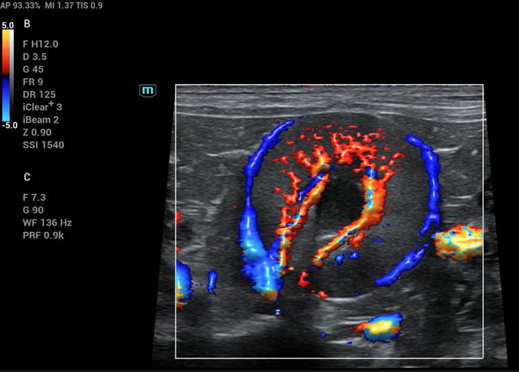
Colour flow doppler in a canine kidney from Mindray Vetus 80
This is what most people think of when you say Doppler and is generally just called Colour Doppler (CF) though can also be referred to as Colour Flow Doppler (CFD) and/or Colour Doppler Imaging (CDI). It is displayed as a colour overlay in a window over a B-mode (or M-mode) image. It is not specific for blood flow, though visualising blood flow is the most common use and detects motion relative to the transducer.
When scanning the abdomen of a panting or otherwise wriggling patient, you will notice colour filling the window (known as flash artefact). This is caused by the motion of the tissues relative to the transducer. Colour Doppler can appear very sensitive to different velocities based on how the settings are adjusted, and there are various artefacts that can impede interpretation if not set correctly. It also has a maximum velocity that can be displayed (Nyquist limit) before artefacts are seen.
As shown in the image, the two colours will appear in the direction of motion relative to the probe. The standard setup is; blue away from the probe, red towards the probe and green, in cardiac imaging, which reflects the degree of flow disturbance present.
Colour Doppler is used widely in small animal and equine practice and becoming increasingly important in farm animal practice.
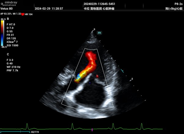
Colour Doppler image from Mindray Vetus 80 showing aortic regurgitation
Power Doppler
Power Doppler image showing renal perfusion
Power Doppler (PD) also known as Power Doppler Imaging (PDI) is very similar to Colour Doppler, except that it is non-directional. Therefore, Power Doppler is not typically used in echocardiography where direction of flow is key to assessing the heart.
Power Doppler offers increased sensitivity, making it easier to visualise small vessels and low-velocity blood flow. It detects the amount of movement rather than direction, so regions with more moving blood cells appear brighter on the image — and unlike other Doppler modes, it has no maximum velocity limit.
The disadvantage is that extra sensitivity means more flash artefact. If the probe or patient is moving, the motion will appear more strongly than with Colour Doppler.
Spectral Dopplers
Pulsed Wave Doppler (PW or PWD) and Continuous Wave Doppler (CW or CWD) are typically displayed as spectral waveforms, with time on the x-axis and velocity on the y-axis. Blood flow is represented as pixels relative to a baseline, flow towards the transducer appears above the baseline, while flow away appears below. The brightness of the waveform correlates with the number of moving red blood cells. Most ultrasound systems can display the B-mode image alongside the spectral trace, although this can reduce the overall image quality of the trace.
Pulsed wave Doppler
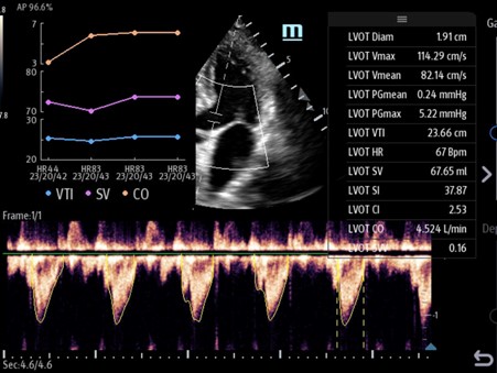
Pulsed Wave Doppler is used primarily in echocardiography but has some applications within abdominal imaging. As the name suggests, Pulsed Wave Doppler uses rapid pulses to measure flow. When activating Pulsed Wave Doppler, you first position the sample volume over the region of interest. This is shown as two horizontal lines on a vertical line.
Pulsed Wave Doppler is region-specific, meaning velocity measurements are taken precisely from the location where the Sample Volume (SV) is placed. However, because it must wait for each pulse to return before sending the next, and due to the speed of sound in tissue, it is limited by a maximum measurable velocity known as the Nyquist limit.
To overcome this limitation, some systems offer a High Pulse Repetition Frequency (HPRF) mode, which sends new pulses before previous echoes have returned. This increases the velocity range but sacrifices exact location accuracy, as multiple sample volumes may appear—indicating that the measured velocity could originate from any of those regions.
Continuous Wave Doppler
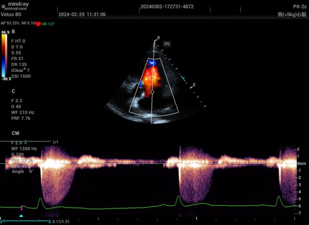
Continuous Waver Doppler image from Mindray Vetus 80
Continuous Wave Doppler lives up to its name by transmitting sound continuously, using two separate crystals in the transducer, one for sending and one for receiving. This continuous transmission allows the system to measure extremely high velocities, essentially without an upper limit, making it ideal for detecting fast blood flow that exceeds the Nyquist limit of Pulsed Wave Doppler.
Unlike Pulsed Wave Doppler, which produces velocity ‘envelopes’ due to its region-specific sampling, Continuous Wave Doppler measures all velocities along the entire sound path. This results in a filled spectral trace that includes both high and low velocities. The trade-off is a loss of spatial specificity, you can’t determine exactly where along the line the flow originates. For this reason, it’s often best to first localise the area of interest using Colour Doppler or Pulsed Wave Doppler before applying Continuous Wave.
Tissue Doppler
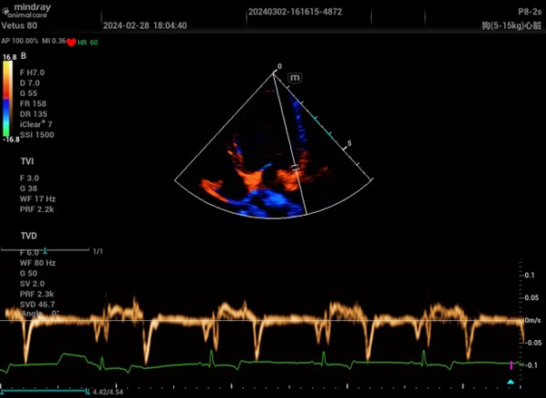
Colour and Pulsed Wave Tissue Doppler Imaging from a Mindray Vetus 80
This mode is used in specialist echocardiography, often called Tissue Velocity Imaging (TVI) or Tissue Doppler Imaging (TDI). There are two types, colour and pulsed wave. They are simply more specialised versions of their namesakes above, optimised for tissue movement rather than blood flow.
Contact us
If you need any assistance with an existing system or purchasing an ultrasound machine don’t hesitate to contact us and one of our experts would be happy to answer any questions you may have. Alternatively, browse through our range of new and refurbished ultrasound products here.