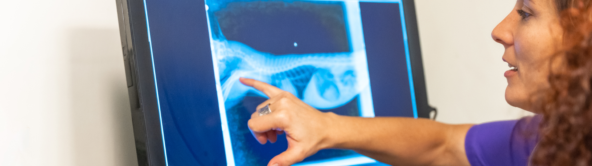
Digital radiography refers to the electronic capture of X-ray images. There are three main technologies behind digital X-ray imaging: CR (Computed Radiography), CCD (Charge-Coupled Device), and DR/DDR (Digital Radiography/Direct Digital Radiography).
X-ray and other technology within healthcare are continuosly evolving and DR systems are a great example of this. Today's DR systems now enable images to be captured and displayed within seconds, allowing a full X-ray series to be completed within minutes. This not only reduces the time animals spend under anesthesia but also improves the overall workflow of your practice. CR systems however are a more manual process, which uses a cassette with a special imaging plate that stores the X-ray image. The cassette must be processed in a separate reader to digitize the image.
If you’re still using a CR (Computed Radiography) system in your veterinary practice, you’re not alone - but you’re also likely feeling the growing pain points: slow workflows, inconsistent image quality, and increasing maintenance needs.
Upgrading to DR (Digital Radiography) isn’t just a technology refresh, it’s a transformation in speed, image quality, and efficiency that directly impacts patient care and practice productivity.
Here’s how DR stacks up against CR and why more practices are making the switch:
1. Speed: Instant Imaging vs. Workflow Bottlenecks
- CR: Requires handling imaging cassettes, running them through a scanner, and waiting up to a minute (or more) per image.
- DR: Provides instant image capture directly to your workstation, usually within 3-5 seconds.
Faster diagnostics, reduced anesthesia time, and quicker emergency workups.
2. Image Quality: Clarity That Matters
- CR: Susceptible to artefacts, scratches, and inconsistent exposure.
- DR: Offers higher resolution, better contrast, and more consistent results, especially for subtle pathologies or low-contrast areas like soft tissue or lungs.
Fewer repeat exposures and more confident clinical decision-making.
3. Workflow: Streamlined vs. Step-Heavy
- CR: Involves multiple manual steps - positioning, scanning, naming files, and uploading.
- DR: Fully digital workflow, often integrated with PACS, reducing human error and saving valuable time
More efficient use of team resources, especially in busy or understaffed practices.
4. Durability and Maintenance
- CR: Imaging plates wear out, need frequent replacement, and require careful handling.
- DR: Flat panel detectors are more robust, require less maintenance, and don’t degrade image quality over time.
Lower long-term costs and fewer equipment headaches.
5. Space and Setup
- CR: Requires a dedicated scanner unit and physical storage for imaging plates.
- DR: Compact and often wireless, making it ideal for smaller spaces or mobile setups.
Reclaim space and maximise your imaging area.
6. Long-Term Value and Future-Proofing
- CR: Older technology with fewer updates and limited support from manufacturers.
- DR: Continuously evolving with better software, AI tools, and integration options.
CR was once a stepping stone into digital imaging, but in 2025, DR is the standard for practices that value speed, quality, and efficiency. Whether you’re a single-site clinic or a multi-location group, switching to DR isn’t just an upgrade, it’s a strategic move for better patient care and a more modern veterinary workflow.
Still using CR? It might be time to talk about what DR could do for you
Contact us
Interested in finding out more? Probo Medical also offer a wide range of solutions for Veterinary Practices. Contact us today for a free product demonstration or quote. Alternatively, browse through our range of X-Ray Veterinary Equipment here.